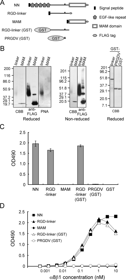
FIGURE 3.
Binding activities of α8β1 integrin to nephronectin and its fragments. A, schematic diagrams of full-length nephronectin (NN) and its fragments. RGD-linker and RGD-linker (GST), the central RGD-containing linker segments expressed in mammalian and bacterial expression systems, respectively; PRGDV, a short RGD-containing peptide modeled after nephronectin and expressed as a GST fusion protein (see Fig. 4A for the peptide sequence). The arrowheads indicate the positions of the RGD motif. B, purified recombinant proteins were analyzed by SDS-PAGE in 7–15% gradient (left and center panels) and 12% (right panels) gels, followed by Coomassie Brilliant Blue (CBB) staining, immunoblotting with an anti-FLAG mAb, or lectin blotting with PNA. The quantities of proteins loaded were: 0.5 μg (for Coomassie Brilliant Blue staining) and 0.1 μg (for blotting with anti-FLAG and PNA) in the left and center panels;1 μg in the right panel. C, recombinant proteins (10 nm) were coated on microtiter plates and assessed for their binding activities toward α8β1 integrin (10 nm) in the presence of 1 mm Mn2+. The backgrounds were subtracted as described in the legend to Fig. 2. The results represent the mean ± S.D. of triplicate determinations. D, titration curves of α8β1 integrin bound to full-length nephronectin (NN, closed squares), the RGD-linker segments expressed in 293F cells (RGD-linker, closed triangles) and E. coli (RGD-linker (GST), open triangles), the MAM domain (MAM, closed diamonds), and the PRGDV peptide expressed as a GST fusion protein in E. coli (PRGDV (GST), open circles). The assays were performed as described in the legend to Fig. 2B. The results represent the means of duplicate determinations.

