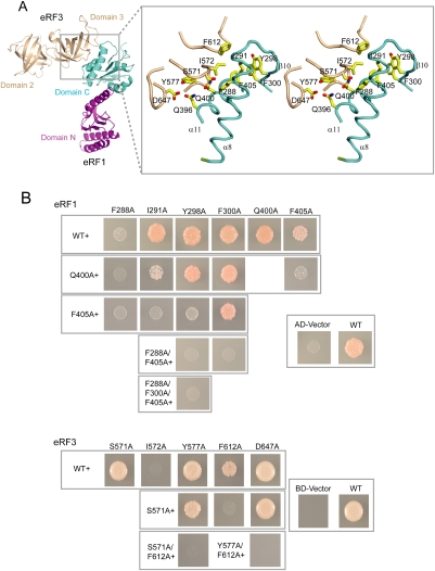
Figure 2.
Interaction between eRF1 and eRF3. (A, left) The SpeRF1/eRF3-23 complex shown as in Figure 1B. (Right) Stereo view of the interface of eRF1/eRF3. Residues involved in the interactions are labeled and shown in yellow stick model. (B) Yeast two-hybrid assays for S. pombe eRF1 and eRF3. (Top) eRF1 wild-type and interface mutations in AD (δ-N2 construct of eRF1) vector against eRF3 in BD vector. (Bottom) eRF3 wild-type and interface mutations in BD vector against eRF1 in AD vector (δ-N2 construct). The first mutation is indicated at the top and additional mutation(s) are indicated to the left in both panels.

