Figure 4.
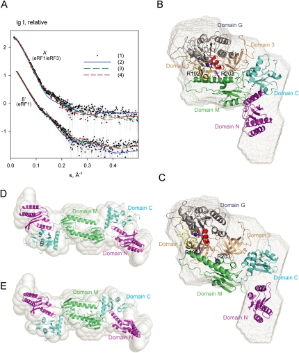
SAXS data and shape reconstructions. (A) Experimental and computed SAXS scattering data. (A′) the eRF1/eRF3/GTP complex, (B′) free eRF1. The logarithm of the scattering intensity is plotted against the momentum transfer s = 4π sinθ/λ, where 2θ is the scattering angle and λ = 1.5 Å is the X-ray wavelength. The plots are displaced along the ordinate for better visualization. (1) Experimental scattering. (2) Computed scattering from the molecular model of eRF1/eRF3 (in A′) and from the crystallographic dimer of eRF1 (in B′). (3) Computed scattering from these two models after rigid body refinement. (4) Computed scattering from the rigid body model of eRF1/eRF3 using extended eRF1 (A′) and from the rigid body model of eRF1 dimer using a compact eRF1 monomer (B′). (B,C) The modeled eRF1/eRF3/GTP complex (B) and the rigid body refined model (C) are superimposed onto the ab initio shape reconstruction (gray mesh). (D,E) The crystallographic dimer of extended eRF1 (D) and the rigid body model of the dimer from extended monomers (E) are superimposed onto the ab initio shape reconstruction (semitransparent beads). The coloring scheme for eRF1 and eRF3 is as in Figure 3E.

