Fig. 6.
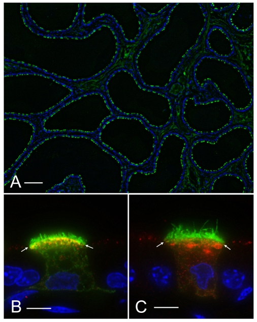
Rat cauda epididymidis perfused in vivo and stained for the V-ATPase B1 subunit (green). Nuclei were stained with DAPI (blue). (A) Numerous B1-positive clear cells were detected. Luminal spermatozoa are absent from these perfused tubules. (B) Higher magnification of a clear cell perfused with a control phosphate-buffered solution adjusted to pH 6.6 and containing the endocytic marker, HRP. Double-labeling for HRP (red) and the V-ATPase B1 subunit (green) was performed. The V-ATPase is distributed between sub-apical vesicles and short microvilli. The yellow staining indicates partial colocalization of the V-ATPase with HRP in endosomes. (C) Clear cell perfused with an `activation' buffer containing bicarbonate and cpt-cAMP. The V-ATPase is mainly located in longer microvilli (green) and no colocalization with HRP-labeled endosomes is detected (red). The staining was performed as previously characterized (Shum et al., 2008). Scale bars, 150 μm (A), 5 μm (B,C).

