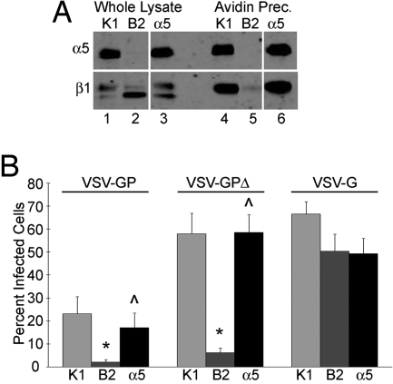
Fig. 1.
Expression of α5β1-integrin enhances EboV GP-mediated infection of CHO cells. (A) Total lysates and surface proteins of CHO K1 (K1), CHO B2 (B2), and CHO B2-α5 (α5) cells were immunoblotted for α5 (Upper) and β1 (Lower) integrin. Panels shown are from the same blot and same exposure; gaps indicate where lanes were removed. (B) The 3 CHO cell lines were infected with VSV-GP, VSV-GPΔ, or VSV-G, and the percentage of infected cells expressing GFP was measured by flow cytometry. Data shown are the averages from 10 experiments. Error bars indicate SEM. *, P ≤ 0.02 relative to CHO K1 cells. ⋀, P ≤ 0.05 relative to CHO B2 cells.

