Fig. 2.
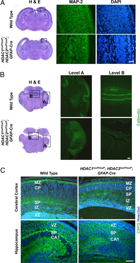
Neuronal defects in HDAC1loxP/loxP; HDAC2loxP/loxP; GFAP-Cre mice. (A) Immunohistochemical staining of MAP-2 on the neocortex of wild-type and mutant mice. Mutant mice show a lack of dendritic extensions compared with control. Scale bar = 40 μm. (B) Immunohistochemical staining for calbindin on wild-type and mutant cerebellum. Mutant cerebellum shows Purkinje cells have failed to migrate and remain among the deep cerebellar nuclei. Scale bar = 40 μm. (C) Immunohistochemistry of CSPGs (green) and Hoechst (blue) of wild-type and mutant cerebral cortex and hippocampus. Scale bar = 40 μm. Deletion of HDAC1 and HDAC2 results in disorganized molecular layers. Hippocampal structures are unidentifiable in mutant mice. MZ, marginal zone; CP, cortical plate; SP, subplate; IZ, intermediate zone; VZ, ventricular zone.

