Fig. 3.
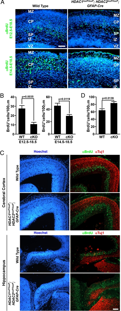
Neuronal birth date and proliferation analysis of HDAC1loxP/loxP; HDAC2loxP/loxP; GFAP-Cre mice. (A) Immunohistochemistry of BrdU (green) and Hoechst (blue) of wild-type and mutant cerebral cortex injected with BrdU at E12.5 or E14.5. Deletion of HDAC1 and HDAC2 results in normal neuronal migration but results in fewer differentiated neurons. (B) Quantification of BrdU+ cells from (A) showing a statistically significant reduction in neurons from double mutant (cko) mice. Scale bar = 40 μm. (C) Immunohistochemistry at E14.5 detecting Hoechst (blue), S-phase neuronal precursors labeled by BrdU (green), and Tuj1 (red) on wild-type and mutant cerebral cortex (Top) and developing hippocampus (Bottom). Scale bar = 40 microns. (D) Quantification of BrdU+ cells at E14.5 to assess proliferation. Deletion of HDAC1 and HDAC2 results in increased proliferation of neuronal precursors at the ventricular zone and reduced number of Tuj1-expressing neurons at E14.5. MZ, marginal zone; CP, cortical plate; SP, subplate; IZ, intermediate zone; VZ, ventricular zone; cKO, double conditional knockout.

