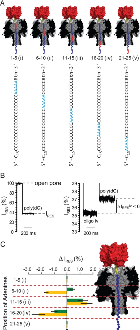
Fig. 2.
Probing DNA recognition by the αHL pore with A5 oligonucleotides. (A) The 5 oligonucleotides (i–v) containing 5 consecutive adenine nucleotides (A5, red circles) at different positions (numbered from the 3′-biotin tag) in an otherwise poly(dC) strand (cytidine nucleotides are shown as blue circles). Only the first 25 of the 40-nucleotide-long sequences are shown. (B Left) The stepwise reduction from the open current value (pore not blocked with DNA) to a residual current (IRES) level of ≈37% when the E111N/K147N pore becomes blocked with a poly(dC) oligonucleotide. (B Right) The IRES levels when a pore is blocked with oligonucleotides of different sequence (oligo iv and poly(dC) are shown). (C) Residual current difference (ΔIRES) between the blockade by oligonucleotides i–v (A) and poly(dC)40 for WT (green bars) and E111N/K147N (orange bars) αHL pores (ΔIRES = IRESi–v − IRESpoly(dC)). The probable location of the adenine (A5) stretch of each oligonucleotide when immobilized with an αHL pore is indicated (Right).

