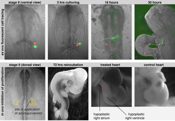
Figure 6.
Panel A shows the movement of fluorescently labeled cells from the caudal splanchnic mesoderm of a stage 9 embryo. The inner mesoderm is labeled with DiO (green) and the outer mesoderm is labeled with DiI (red). For a spatial appreciation of the inner and outer mesoderm refer to Figure 4 (indicated with ▲ and △, respectively). With culturing the outer mesoderm can be seen to be incorporated into the inflow of the heart, while the inner mesoderm moves, via the dorsal pericardial wall, into the outflow of the heart. Panel B shows the effect of local inhibition of proliferation. At the right side of the embryo, a focus in the caudal and inner mesoderm was exposed to Aminopurvanolol, dissolved with DiI. After reincubation 9 out of 16 treated embryos suffered from hypoplasia of both the right ventricle and the right atrium. Of the other embryos, 2 deceased before analysis, 1 showed only outflow-malformations, and 4 seemed to be unaffected.

