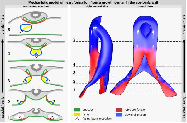
Figure 7.
illustrates heart formation from the proliferating growth-center in the dorsal pericardial wall. The left column shows transverse sections, ranging from cranial in an old embryo to caudal in a young embryo. Firstly, outer mesoderm luminizes and stops to proliferate. Next, it bends inwards and fuses to form the ventral wall of the heart tube (2-4). The inner mesoderm keeps proliferating and forms the pericardial back wall and its connection with the heart tube (5). These sections were transformed into the model that is shown on the right. The model shows that expansion from the caudal growth-center leads to a radial addition of medially located mesoderm to the heart tube. After regression of the dorsal mesocardium, addition to the arterial pole occurs via the pericardial back wall.

