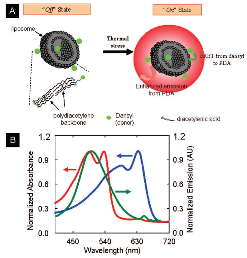
Figure 2.

(A) Schematic representation of a polydiacetylene liposome prepared with a mixture of the fluorescent diacetylene 1 and PDA. The “Off” state represents when FRET efficiency (E) from dansyl to PDA is low, and the “On” state represents when E is large. (B) Normalized absorption spectra of blue (blue curve) and red forms (red curve) PDA and emission spectrum of dansyl fluorophores (green curve) attached to PDA liposomes (λex = 337 nm). The contribution of PDA direct excitation to the total PDA emission for λex = 337 nm is extremely small.6a
