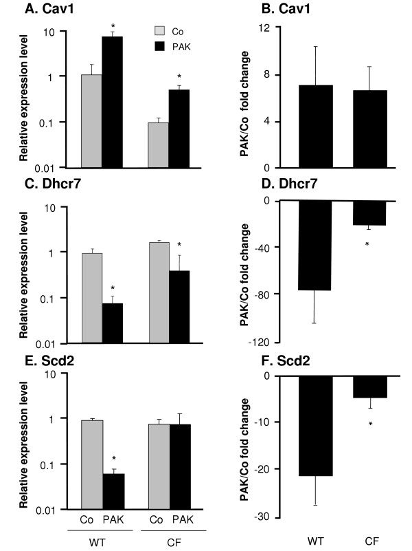
Figure 5.
Confirmation of microarray results by real-time RT-PCR. DC from WT and CF mice were infected in vitro with P. aeruginosa for 4 h. RNA levels for three genes were measured by quantitative real-time RT-PCR. Relative expression levels in the samples were calculated using the ΔΔCt method, with GAPDH as internal normalization control. A, C and E. The y-axis represents the relative gene expression level for Cav1, Dhcr7 and Scd2 in the uninfected control DC (gray) and P. aeruginosa infected DC (black). B, D, and F. The y-axis represents fold change of Cav1, Dhcr7 and Scd2 expression upon P. aeruginosa infection compared to the control in both groups. Shown are the means ± SEM of three pairs of DC samples from WT and CF mice with or without P. aeruginosa infection. *denotes p < 0.05.

