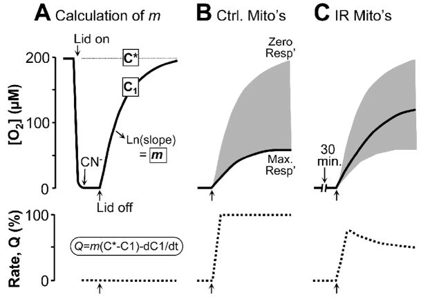
Figure 2. Theoretical principles of application of open-flow respirometry to hypoxia-reoxygenation of isolated mitochondria in a Clark O2 electrode chamber.
Mitochondria are incubated in respiration buffer + respiratory substrates & ADP, in a respiration chamber fitted with an O2 electrode. (A): Where indicated, the lid is closed and mitochondria respire to bring [O2] to zero (i.e. hypoxia). 1mM CN− is then added to block respiration and the lid is opened. The resulting rise in [O2] is due to O2 diffusion into the chamber, and equilibrium with room air is eventually obtained. The first-order rate constant for O2 diffusion (from Ln[O2] vs. time) is the mass transfer coefficient, m. C* is the [O2] of air saturated buffer alone (~200μM). C1 is the [O2] measured by the O2 electrode (solid line). Respiration rate (Q, lower dotted trace) is zero throughout “reoxygenation”, due to the CN− inhibition. (B): Mitochondria are incubated as in panel A, without CN−. Opening the lid results in a slower rise of [O2] (C1) due to the equilibrium between O2 diffusing in and O2 consumed by the mitochondria. The shaded gray area represents the range of possible [O2] recovery curves, resulting from zero or maximal respiration. Respiration rates (Q) are calculated instantaneously during reoxygenation, using the equation Q=m(C*−C1)/dC1/dt. (C): Mitochondria are incubated as in B, but with hypoxia. Respiration (Q) recovers rapidly at reoxygenation, but Ox-Phos damage prevents later full recovery.

