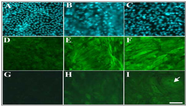
Figure 2.
FGF- and vitreous-induced phosphorylation of Akt and ERK1/2 during lens fibre differentiation. Immunofluorescent labelling of β-crystallin (D, E, F) or γ-crystallin (G, H, I) in lens explants cultured without growth factors (A, D, G), with 100ng/ml FGF (B, E, H) or with 50% vitreous (C, F, I). Cell nuclei were counterstained with Hoechst dye (A, B, C). Cells in control explants remain cuboidal in shape and do not express β– and γ-crystallin (D, G). FGF- and vitreous-treated cells elongate and accumulate β– (E, F) and γ-crystallin (H, I). Vitreous-induced fibre differentiation results in the presence of larger cells (I, arrow). Scale bar, 50 μm.

