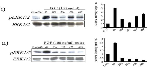
Figure 8.
Comparison of ERK1/2 phosphorylation profiles induced by a high dose of FGF (100 ng/ml) present throughout the culture period (i) or for a defined period of time (ii, FGF pulse treatment). (i) Representative western blot of lens explants cultured with no growth factors or with FGF for 6h, 30h, 36h, 42h and 48h. Elevated ERK1/2 phosphorylation was detected well after 6h, with high levels still detected at 42h. (ii) Representative western blot of lens explants cultured with no growth factors or with a 30min pulse treatment of FGF and assayed after 6h, 30h, 36h, 42h and 48h. Elevated ERK1/2 phosphorylation was detected at 6h, and dropped to basal levels throughout the remainder of the culture period.

