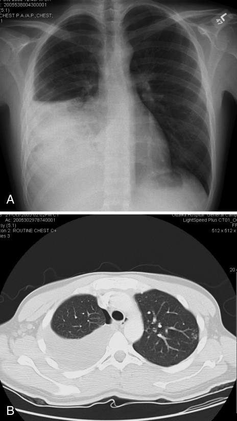
Figure 2).
A A plain chest radiograph showing a moderate-sized right pleural effusion; B Computed tomography of the thorax in the lung window settings confirmed a moderate-sized right pleural effusion. Consolidative changes of the lateral segment of right middle lobe and right lower lobe were also seen. A small pneumothorax was seen following a thoracentesis. Centrilobular nodules in a tree-in-bud pattern at the apicoposterior segment of left upper lobe were present

