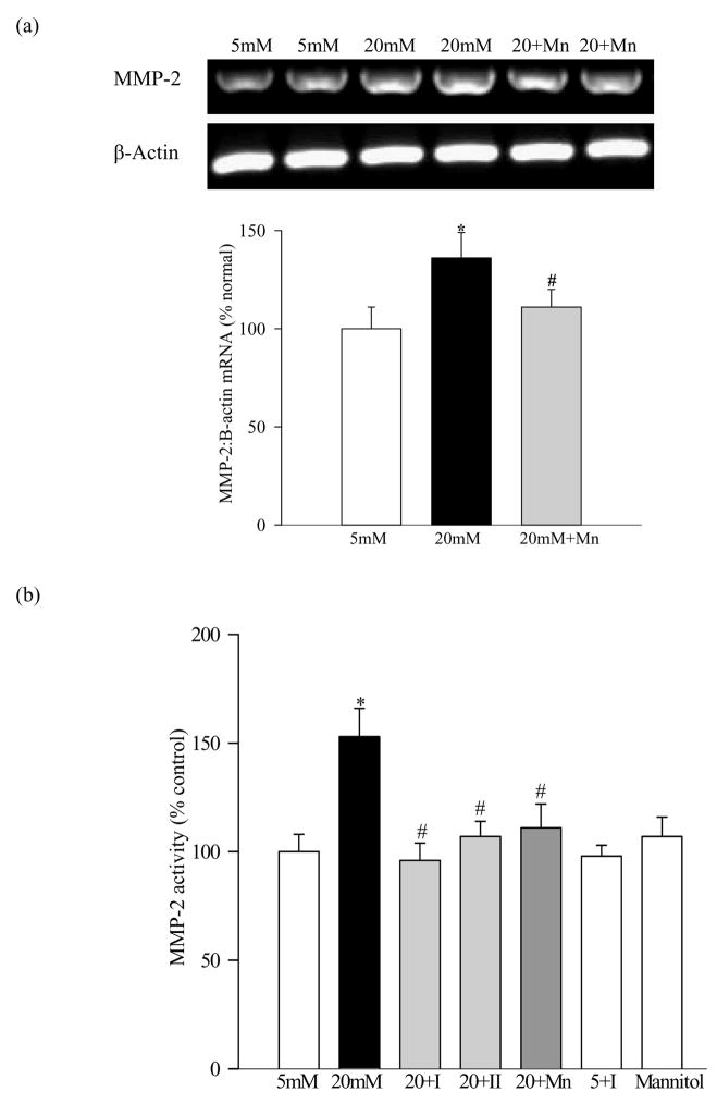
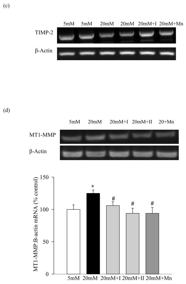
Figure 1.
Effect of high glucose on MMP-2 and its regulators in retinal endothelial cells: Bovine retina endothelial cells from 4–7th passage were incubated in 5mM glucose or 20mM glucose medium for 4–5 days in the presence or absence of 10μM MMP-2 inhibitor I or 5μM MMP-2 inhibitor II, or 200μM MnTBAP. At the end of incubation medium was collected, and the cells were washed with phosphate buffer saline and collected. (a) Gene expression of MMP-2 and that of β-actin in the cells was determined by semi-quantitative PCR using the primers given in table I. (b) The gelatinase activity of MMP-2 was determined in the medium. (c) and (d) The levels of mRNA of TIMP-2 and MT1-MMP were determined by semi-quantitative PCR respectively, and were adjusted to the mRNA levels of β-actin in each sample. Each measurement was made in duplicate in at least three different cell preparations. The values obtained from the cells incubated in 5mM glucose are considered as 100% (control). *P<0.05 compared to the values obtained from the cells incubated in 5mM glucose, and #P<0.05 compared to 20mM glucose. 20+I= 20mM glucose+10μM MMP-2 inhibitor I; 20+II=20mM glucose+5μM MMP-2 inhibitor II; 20+Mn=20mM glucose+200μM MnTBAP; 5+I=5mM glucose+10μM MMP-2 inhibitor I


