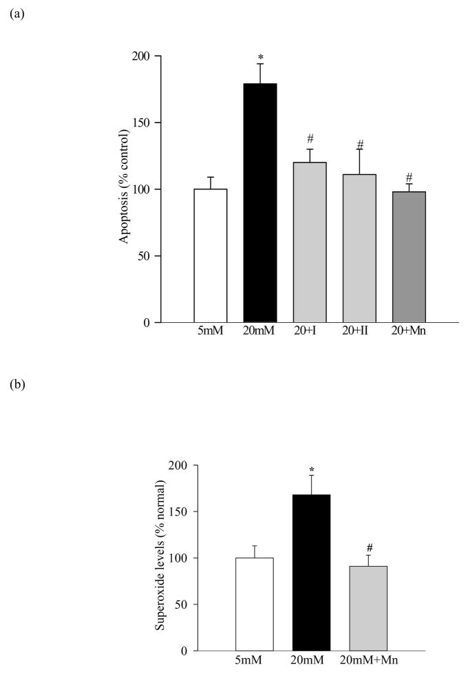
Figure 2.
Effect of inhibition of MMP-2 and MnTBAP on the apoptosis of retinal endothelial cells: (a) Apoptosis was measured by performing ELISA for cytoplasmic histone-associated-DNA-fragments using an assay kit from Roche Diagnostics. The graph shows the values obtained from the cells incubated with glucose for five days, and these values were adjusted to the total DNA. (b) Superoxide levels were measured in the mitochondria by lucigenin method. The values obtained from the cells incubated in 5mM glucose are considered as 100%. Each measurement was made in duplicate in at least four different cell preparations. *P<0.05 compared to the values obtained from the cells incubated in 5mM glucose, and #P<0.05 compared to 20mM glucose.

