
Figure 4. Cholesterol content of ex vivo biotinylated platelets after infusion into WT or SR-BI KO recipients.
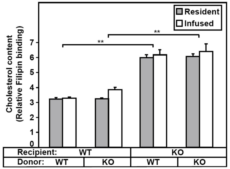
Platelets from WT or SR-BI KO mice were labeled with biotin and infused into WT or SR-BI KO recipients. Next day the recipient mice were bled, the platelets stained with GPIIbIIIa-APC, PE-SA and filipin, and the cholesterol content (filipin staining) of resident (non-biotinylated) and infused platelets was determined by flow cytometry. (n=3–4). The data are from one of two similar independent experiments (**P<0.001).