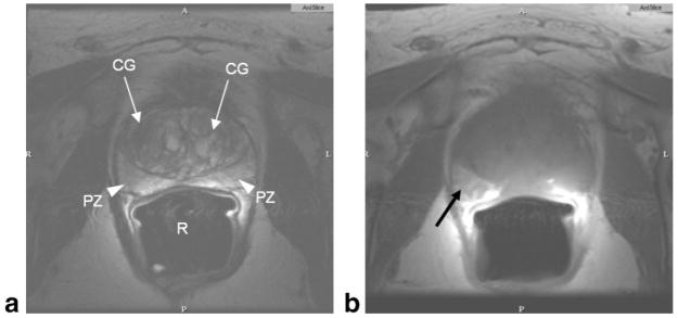
Figure 2.
Normal prostate MRI. Axial T2W (a), and axial T1W (b) image of prostate taken at 1.5T using an endorectal coil. Central gland (CG, white arrows) in the T2W image shows benign prostate hyperplasia. Peripheral zone is indicated as PZ (white arrowheads). Air in the rectum (R) is due to the endorectal coil balloon. Hyperintense region of the right peripheral zone in the T1-weighted image (black arrow) indicates hemorrhage.

