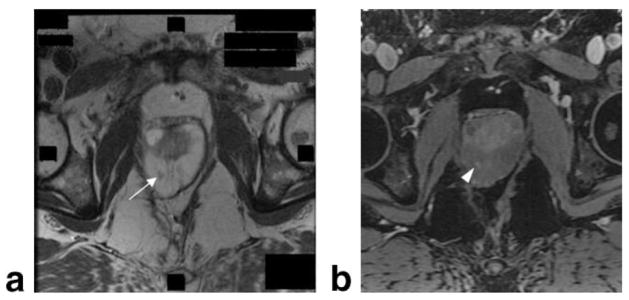
Figure 5.
MR of the prostate at 3T. Axial MRI of the prostate obtained prior to biopsy at 3T using a body coil. Prior rectal surgery precluded the use of an endorectal coil. Within T2W image (a), low signal region (white arrow) indicates a focal lesion in the right mid-gland. T1-weighted image (b) shows postgadolinium contrast enhancement of the lesion (white arrowhead).

