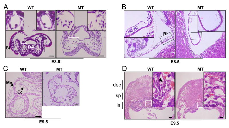
Figure 6. Histology of Er71 mutants.
(A, B) H & E staining of E8.5 embryos. The boxed area is shown at higher magnification. Bl, blood island; DA, dorsal aorta. Scale bars; 50μm. (C) Sagittal sections of wild type and Er71−/− hearts from E9.5 embryos. Ec, endocardium; Mc, myocardium. Scale bars; 50 μm. (D) Sagittal sections of the placenta of E9.5 embryos. Higher magnification of the labyrinth layers of the wild type and Er71−/− placentas is shown in the box. dec, maternal decidual tissue; sp, spongiotrophoblast layer; la, labyrinth layer. Scale bars; 200 μm.

