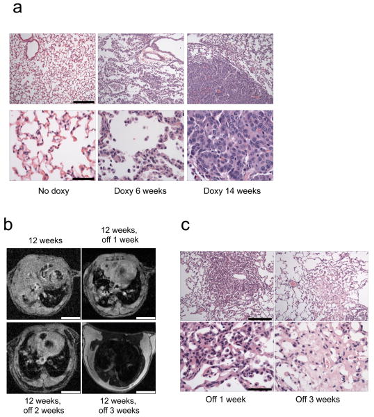
Figure 1. Development of a Tet-inducible PIK3CA H1047R mouse model of lung tumorigenesis.
(a) Histological analyses of lungs derived from the bitransgenic inducible Tet-op-PIK3CA H1047R /CCSP-rtTA (line #13) mice. Lungs from mice not induced with doxycycline, or those from mice induced for 6 and 14 weeks are shown. Adenocarcinoma is present in the lungs of mice induced with doxycycline after 6 and 14 weeks, respectively. Scale is 200μM and 50μM for upper and lower panels respectively. (b) Rapid disappearance of lung tumors following withdrawal of doxycycline. PIK3CA H1047R /CCSP-rtTA mice were placed on a doxycycline diet for 12 weeks to induce tumor formation, and tumors were assessed by MRI. The same mice were then taken off doxycycline and re-imaged 1, 2 and 3 weeks later. A representative example is shown. Scale is 4.5 mm. (c) Histological analysis of lungs after doxycycline withdrawal. PIK3CA H1047R /CCSP-rtTA mice were placed on a doxy diet until tumors were confirmed by MR imaging. Doxycycline was then withdrawn from their diets, the mice were sacrificed, and their lungs were examined histologically. Shown are the histology sections from two different mice after doxy withdrawal for 1 and 3 weeks respectively. Scale is 200μM and 50μM for upper and lower panels respectively.

