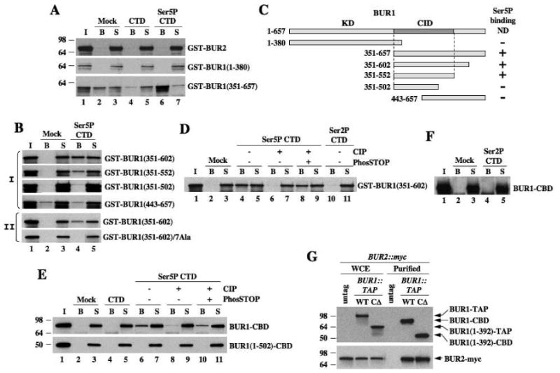
Fig. 2. BUR1 binds to Ser5P-CTD peptides.

(A-D) Biotinylated CTD peptides (1.5μg) phosphorylated on Ser5 (Ser5P-CTD) or unphosphorylated (CTD) were adsorbed to streptavidin-coated magnetic beads. Recombinant GST-BUR2 or GST-BUR1 fragments were incubated with beads alone (Mock) or beads bearing peptides at 4°C. Bound (B) and unbound proteins in supernatant (S) were subjected to SDS-PAGE and Western analysis with myc antibody. (C) Schematic summarizing results in (A-B). (E-F) BUR1-CBD and BUR1(1-502)-CBD purified from yeast were used for peptide binding assays as above and detected using anti-TAP antibodies. In (D-E), immobilized peptides were treated with CIP in the presence or absence of PhosSTOP prior to incubation with proteins. (G) BUR1-TAP and BUR1-CΔ-TAP were purified from BUR2::myc strains and subjected to SDS-PAGE and Western analysis, along with the starting extracts (WCE), using antibodies against TAP (upper panel) and myc (lower panel).
