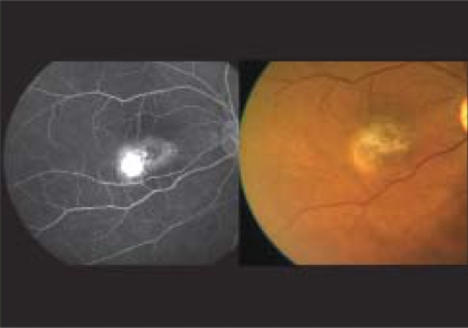
Figure 1.
Color fundus picture (right) reveals perifoveal retinal opacification, refractile crystalline deposits and SRNVM temporal to the fovea along with ILM striae suggestive of proliferative Type 2 idiopathic macular telangiectasia. Fundus fluorescein angiography (left) reveals intense leakage from the SRNVM

