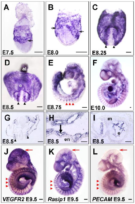
Figure 1. Expression of Rasip1 in vascular endothelium during early embryogenesis.

In situ hybridization showing expression of Rasip1 in embryonic vessels at stages indicated (A-I, K). A-F) Whole mount in situ hybridization showing whole stained embryos. Note expression in both scattered angioblasts (thin arrows), forming dorsal aortae (black arrowheads), intersomitic vessels (ISVs) (red arrowheads) and yolk sac vessels (thick arrows). G-I) Transverse sections of in situ hybridizations showing endothelial-specific expression of Rasip1 in G) dorsal aortae, H) yolk sac vessels, and I) heart endocardium. J-L) Comparison of Rasip1 expression with that of the vascular markers VEGFR2 and PECAM, in E9.5 embryos. Note overall similarity of expression, especially in the ISVs (red arrowheads) and trunk vessels. Note difference in intensity of vascular staining in distinct regions, such as the cephalic vessels (red arrows). al, allantois; e, endocardium; en, endoderm; m, myocardium. The scale bars represent 200μm in all panels except J-L, where they represent 50μm.
