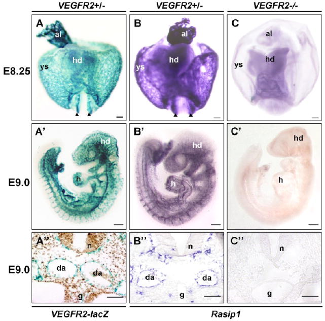
Figure 3. Expression of Rasip1 is restricted to VEGFR2-dependent endothelium.

Whole mount in situ hybridization and β-galactosidase staining to detect expression of Rasip1 and VEGFR2. A,B,A’,B’) Comparison of Flk1(VEGFR2)-lacZ staining and Rasip1 expression. Note overall similarity of expression. Rasip1 expression closely resembles Flk1(VEGFR2)-lacZ expression at both E8.5 (A,B) and E9.0 (A’,B’). C,C’) VEGFR2-/- null embryos, lacking all endothelium. Embryos in C-C” have been stained by in situ hybridization for Rasip1 expression and allowed to develop same length of time as wildtype embryos in B-B”. Note complete lack of Rasip1 expression in these mutants. A”-C”) Sections through embryos in A’-C’ showing presence of aortae and perineural vascular plexus in wildtype embryos, while these vascular structures are missing in VEGFR2-/- embryos. al, allantois; g, gut tube; h, heart; hd, head; da or black arrowheads, dorsal aortae; n, neural tube; ys, yolk sac. The scale bars represent 200μm (A’-C’) and 50 μm (A-C, A”-C”).
