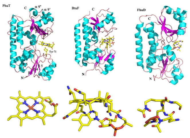
Figure 9.
Structure of PhuT compared with BtuF and FhuD. The major elements of the secondary structure according to the BtuF structure are shown on the PhuT structure. The ligands for each protein are also shown. The PDB accession code for BtuF and FhuD are, 1N2Z and 1K7S, respectively (modified from [48]). Figures were made with PyMOL [162].

