
Fig. 3.
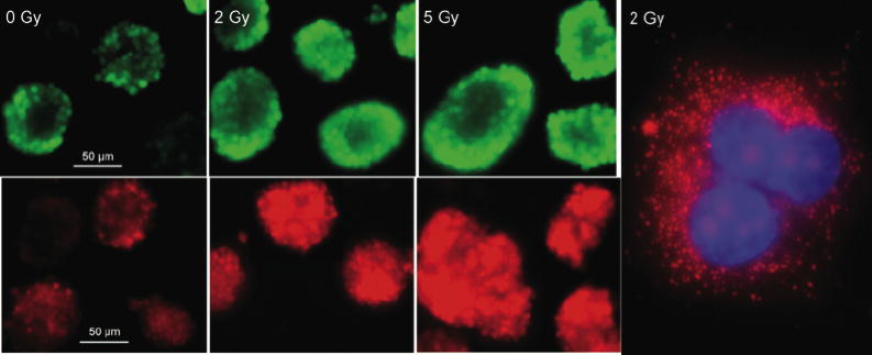
Imaging oxidative stress in living multipotent neural precursor cells. Neural precursor cells received either 0, 2 or 5 Gy and were incubated 1 day later with the carboxy-methyl diacetate analog of reduced fluorescein (CM-H2DCFDA) (upper panels), or Mitosox (lower panels). Cell were rapidly imaged at 60X to prevent photobleaching. Irradiation induced increased staining with both dyes, when compared to cells that were unirradiated. Neural precursor cells were irradiated with 1 Gy and 1 day later, superoxide output from mitochondria the was detected using Mitosox staining (red) and confocal microscopy (right panel). Nuclear counterstaining was done with DAPI.