
Fig. 8.
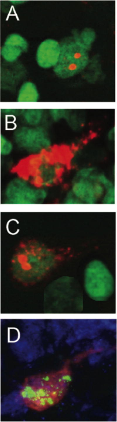
Qualitative characterization of Arc expression in the granule cell layer neurons of the dentate gyrus. Arc was induced by 2 five minute behavioral explorations of a novel environment that were separated by 25 minutes. Intranuclear foci of Arc mRNA were induced by the second exploration, ~ 5 minutes before tissue collection, and were detected using fluorescent in situ hybridization (A). Cytoplasmic Arc mRNA (B) and Arc protein (D) could be detected in neurons activated by the first exploration. Both nuclear foci and cytoplasmic Arc mRNA were seen in ~ 90% of cells immunoreactive for Arc (C). In animals given BrdU to label newly born neurons, labeling with antibodies against BrdU (green) and Arc protein (red) showed dual labeling, indicating that newly born neurons were functionally integrated into the dentate gyrus (D). Digoxine labeled Arc antisense probe was detected with Cy3 (red, A-D), and immunofluorescence staining detected Arc protein (D). Cell nuclei were counterstained green or blue and the magnification for all the images was 63X.