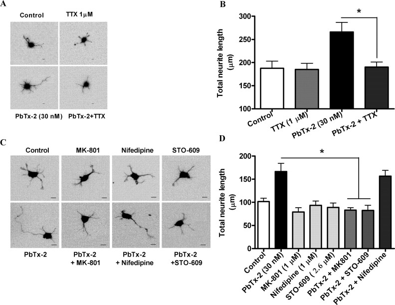
Figure 10.
Pharmacological evaluation of signaling pathways involved in PbTx-2-induced neurite outgrowth. A, Representative images (scale bar, 10 μm) and respective quantification (B) of neurite extension at 24 h. The cerebrocortical neurons were treated with 30 nm PbTx-2 in the presence and absence of 1 μm TTX at 3 h after plating. Bars represent mean ± SEM of 30 cells. *p < 0.05, unpaired t test. C, D, Representative images (scale bar, 10 μm) (C) and quantification (D) of neurite extension at 24 h after plating. The 30 nm PbTx-2 exposure was examined in the presence or absence of MK-801 (1 μm), STO-609 (2.6 μm), or nifedipine (1 μm) beginning at 3 h after plating. Each bar represents the mean ± SEM of 30 cells. PbTx-2-enhanced neurite extension was significantly blocked by MK-801 and STO-609 (*p < 0.05, ANOVA followed by Dunnett's multiple comparison test).

