
Figure 1. Lgd is a novel cytoplasmic C2 domain protein.
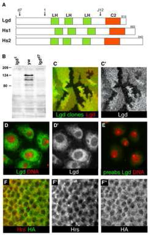
(A) Schematic representation of the protein structures of Drosophila lgd and the two human homologs. The arrow labeled ‘d7’ indicates the position at which the lgdd7 allele has a frameshift mutation: the removal of a single nucleotide results in a new translational frame and a premature stop codon after the addition of 22 new amino acids. The arrow labeled ‘1’ indicates the position at which the lgd1 allele has a frameshift mutation: two nucleotides are removed resulting in a new translational frame and a premature stop codon after the addition of 15 new amino acids. The arrow labeled ‘J12’ indicates the position at which the EMS-induced allele, lgdJ12, contains a nonsense mutation: Q626 is changed to a stop codon. Green boxes indicate the Lgd homology domains (LH), and orange boxes indicate C2 domains. (B) Western blot to detect Lgd protein in extracts from lgd1, yw, and lgdd7 third instar larvae. The lgd1 and lgdd7 alleles truncate the protein N-terminally to the epitope used to generate the Lgd antibodies. The band at approximately 120kD contained the majority of Lgd protein, and was missing in both of the lgd mutant lanes. Other specific bands of lower molecular weight were also occasionally seen. (C–C′) Lgd antibodies specifically detect Lgd protein in imaginal discs. lgd mutant clones, indicated by the lack of GFP (green), stained with α–Lgd antibodies (red in C and gray in C′). (D–E) S2 cells stained with Lgd antibodies (green in D and E and gray in D′) and Topro (red). (E) Lgd antibodies incubated with Lgd antigen prior to use for staining. The preabsorbed Lgd antibodies (green) did not display specific staining, demonstrating that the Lgd antibodies specifically detect the Lgd antigen. (F–F″) C5 Gal4 driven UAS-HA-Lgd in the wing pouch of third instar imaginal discs, detected by α-HA (green in F and gray in F″). HA did not specifically colocalize with α-Hrs (red in F and gray in F′).