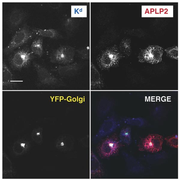
Figure 4.
The Golgi is the large compartment in which Kd and APLP2 co-localized. APLP2-FLAG, etKd, and YFP-Golgi were transiently transfected into HeLa cells. The cells were fixed and then incubated with anti-Kd antibody 34-1-2 and rabbit anti-FLAG and fluorescently labeled secondary antibodies in staining solution. Images were analyzed with a Zeiss LSM 5 Pascal confocal microscope. Blue = folded Kd; red=APLP2; yellow = YFP-Golgi; white = co-localized APLP2 and Kd (fluorescence from Kd, APLP2, and YFP-Golgi, individually, is shown in black and white in the figure). Bar, 10 μm.

