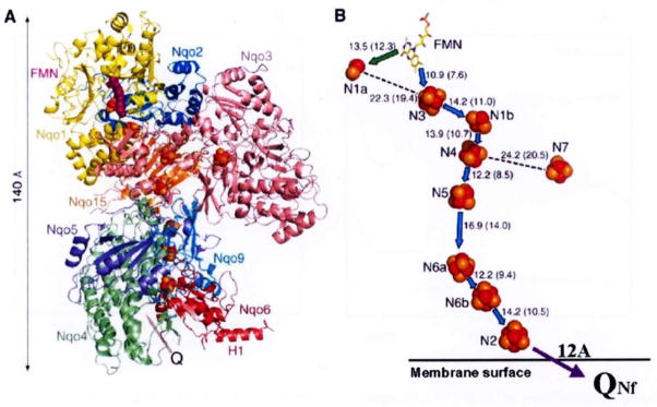
Fig. 1.
Architecture of the hydrophilic domain of Thermus thermophilus HB-8 NADH-Q oxidoreductase (complex I). A, side view, possible quinone binding site (Q) is indicated by an arrow. B, arrangement of redox centers, (by Sazanov and Hinchliffe (2006) Science 311, 1430). Location of the protein-bound QNf (which is shown here in reference to cluster N2) was calculated based on our spin-spin interaction data [35].

