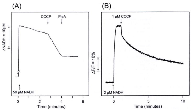
Fig. 4.
(A) The measurement of NADH oxidation (340–420nm). Proteoliposomes (containing 0.003 mg complex I and 0.15 mg phospholipids) were suspended in an 1 ml buffer solution (15 mM Na2SO4, and 5 mM HEPES; pH 7.5) and the suspension was incubated with 100 μM DBQ for 3 minutes. Concentrations of added substances: 50 μM NADH, 0.4 μM CCCP, and 0.5 μM piericidin A (Pie. A). Temperature: 30°C. (B) Fluorescence measurement (excitation 595 nm, emission 615 nm) of the formation of an inside-positive membrane potential with electron transport. The potential was formed with the addition of 2 μM NADH, but it was destroyed by 1 μM gCCCP. The suspension contained 0.003 mg complex I/ml, 2 μM DBQ, 2μM DiBAC4,(5), 15mM Na2SO4, and 5 mM HEPES (pH 7.5). Temperature: 25°C.

