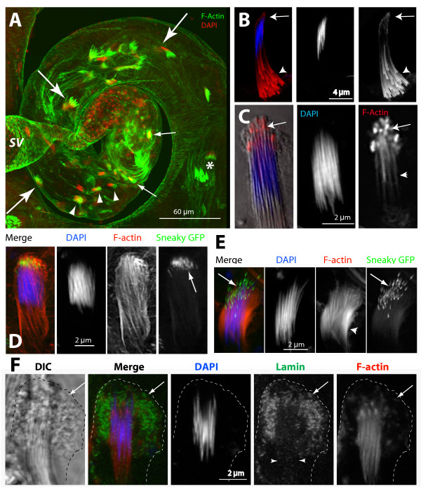
Figure 1.
F-actin-rich structures cap the mature nuclei at the beginning of sperm individualization. (A) Wild-type testis stained with fluorescein isothiocyanate (FITC):phalloidin (green) and 4',6-diamidino-2-phenylindole (DAPI) (red), respectively. Spermatid NBs associated with investment cones (*), at the early (arrows) and intermediate (arrowheads) stages of individualization, and of the coiled-up stages (fine arrows) are marked. Rostral ends of the NBs moved very slowly towards the SV; see Additional files 1 and 2. (B), (C) DAPI (blue) and rhodamine isothiocyanate (RITC):phalloidin (red) stained isolated cysts show F-actin organization at the rostral ends of spermatids during individualization. (B) Arrow indicates F-actin (red) accumulation at the rostral ends of spermatids at the beginning of the investment cone (arrowhead) assembly. (C) Overlay of the differential interference contrast (DIC) picture (gray scale) of the isolated NB containing the needle shaped nuclei (blue) and the F-actin (arrows) cap at the rostral ends. (D), (E) Isolated cysts from sneaky-GFP (green) testis strained as above. (D) NB of a post individualized spermatids. Actin caps appeared around the acrosomes marked by sneaky-GFP (arrows). (E) NB of a relatively later stage bundle (post coiling) found at the testis base. F-actin staining disappeared from the rostral ends but increased laterally (arrowhead). This is presumed to be the penultimate stage before the sperm is released. (F) Combined DIC (Grey) and epifluorescence image of the head cyst cell and associated spermatids stained with anti-Lamin (Dm0) (green), DAPI (blue) and RITC: phalloidin (red). The cell perimeter (broken line) and Lamin-rich membrane folds inside the cell (arrows) are marked. Lamin also localized along the F-actin extensions (arrowheads).

