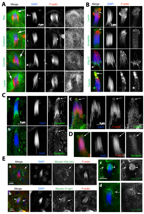
Figure 4.
Immunohistochemical characterization of the actincap. (A) Confocal images of head cyst-cells in intact testis containing mature and individualized sperm nuclei from the basal regions of testes, stained with: ras, DE-cadherin, crumbs and lamin antisera (all in green), as well as rhodamine isothiocyanate (RITC): phalloidin (red) and 4',6-diamidino-2-phenylindole (DAPI) (blue). Arrows indicate staining in the membranous folds inside the head cyst cell and arrowheads indicate the head cyst cell nuclei. (B) Isolated mature spermatids without the head cyst cells as found in the testis squash preparations stained with lamin (Dm0), armadillo, tubulin and ERp72 antisera (indicated at the left side panel of each set). (C), (D) Similar preparations stained with the dynamin (a), syndapin (b) and WASP (c) as well as (D) Syntaxin (green). Arrows indicate positions of the actin caps. (E) Head cyst cells inside testis immunostained with the (a) ck/myosin VII and (b) jar/myosin VI, respectively, or expressing (c) the sqh-GFP and (d) the clathrin light chain:GFP (clc-GFP ), respectively. (a) ck (green) staining (arrows) coincided with the membranous fold and (b) anti-jar stained the actin cap (arrows) as well as a few punctate spots in the cytoplasm (arrowheads). (c) The sqh-GFP localized along the actin caps (arrowheads) and in punctate spots (arrows) in the cytoplasm. (d) clc-GFP mostly localized in the punctate spots (arrows) in the cytoplasm. The sqh-GFP transgene is expressed through its own promoter in sqh homozygous mutant background and the UAS-clc-GFP is expressed by using the actin5CGal4 (see Additional file 6 for details).

