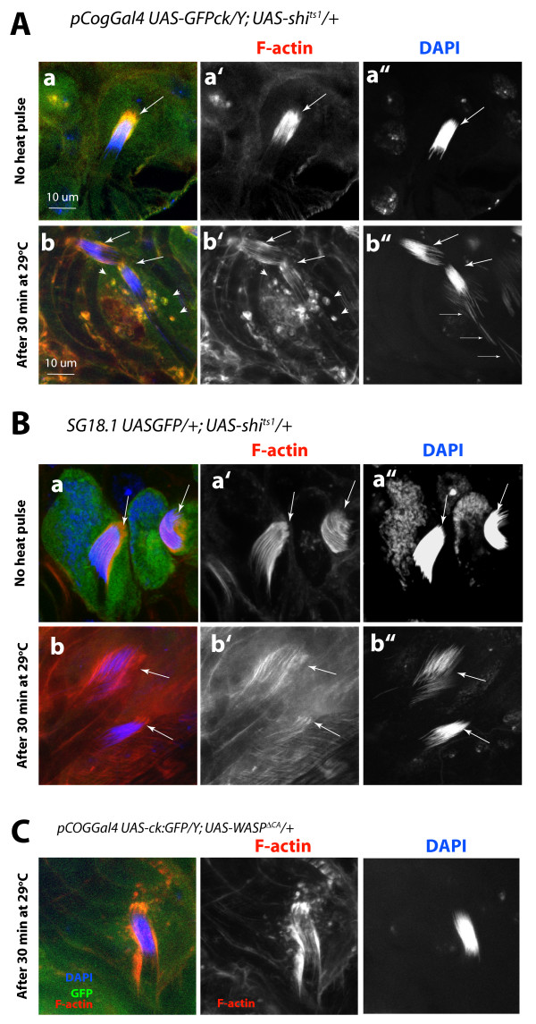
Figure 8.
Transient disruption of the shibire function in the head cyst cell caused actin cap and nuclei bundle disruptions. The UAS-shi ts1 expression in the head cyst cells due to the (A) pCOGGal4 and (B)SG18.1Gal4 drivers, respectively, disrupted the actin caps and nuclei bundles (NBs) at non-permissive temperature (29°C). Rhodamine isothiocyanate (RITC):phallidin and 4',6-diamidino-2-phenylindole (DAPI) staining of these testes (a) before and (b) after the heat pulse showed visible disruptions of the actin caps and NBs (arrows). In addition, there was punctate accumulation of F-actin in the head cyst cell cytoplasm (arrowheads) after the heat pulse. Fine arrows indicate mature nuclei separated from the actin caps. This is not found in the wild-type controls treated in a similar manner. The actin cap and the NB morphology remained unaltered even after 30 minutes at 29°C in the pCOGGal4 UAS-GFPck/Y and SG18.1 UAS-GFP/UAS-actin:GFP testes (data not shown). (C) The UAS-WASP ΔCA expressions by using the pCOGGal4 caused no detectable NB disorganization and a mild accumulation F-actin rich spots in the cytoplasm after a 30 minute heat pulse. (See Additional file 5 for further details.)

