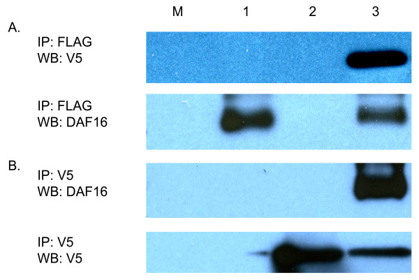
Figure 3.
Co-immunoprecipitation of FTT-2 and DAF-16 expressed in human embryonic kidney 293 cells. Cells were transfected singly (4 μg) or co-transfected (2 μg each) with pcDNA3.1V5/Ac-ftt-2 and pCMV4FLAG/Ac-daf-16. At 16 hrs, the cells were incubated with 20% serum, and lysates prepared 24 hrs later. Lane M, cells transfected with empty pcDNA3.1/V5-His vector (mock); lane 1, cells transfected with Ac-daf-16 alone; lane 2, cells transfected with Ac-ftt-2 alone; lane 3, serum treated co-transfected cells. A. Immunoprecipitation with anti-FLAG (M2) agarose. Top panel, Western blot with anti-V5 antibody; bottom panel, the same blot stripped and probed with DAF-16 antiserum. B. Immunoprecipitation with anti-V5 agarose. Top panel, Western blot with DAF-16 antiserum; bottom panel, the same blot stripped and probed with V5 antibody.

