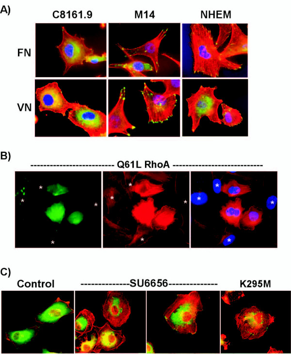
Figure 3.
Src activation prevents RhoA-dependent formation of stress fibers and focal adhesions. A) C8161.9, M14, or normal cultured melanocytes (NHEM) were plated on fibronectin (FN) or vitronectin (VN) for 2 hours and the extent of stress fiber formation was monitored by staining with phalloidin (red). Focal adhesion formation was monitored by immunostaining with paxillin antibodies (green). Cells were counterstained with Hoescht to detect nuclei (blue). B) C8161.9 cells were transiently transfected with GFP expressing plasmid and an active form of RhoA (Q61L). Cells were plated on vitronectin for 2 hours and stained with phalloidin (red) to monitor actin structures in GFP-positive transfected (green) cells. Only the three cells expressing green GFP (i.e. transfected with RhoA (Q61L)) displayed prominent stress fibers. The nuclei of cells not expressing GFP/RhoA Q61L are marked by asterisks. C) C1861.9 cells were left untreated (control), treated with SU6656, or infected with dominant interfering mutant Src (K295M) virus. Cells were plated on vitronectin for 2 hours and stress fiber formation monitored by staining with phalloidin (red) and focal adhesion formation by immunostaining with paxillin antibodies (green).

