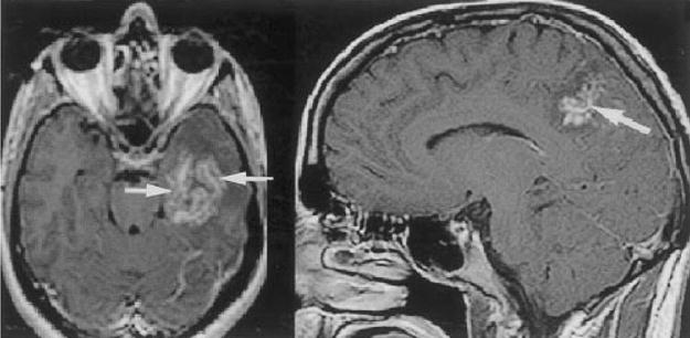
Figure 4.

Two different patients with cerebral schistosomal infection. Contrast-enhanced T1-weighted axial and sagittal images demonstrate central linear enhancement surrounded by multiple enhancing punctate nodules, forming an “arborized” appearance (arrows). Pathologically, this pattern correlated with a host granulomatous response to Schistosoma spp. ova. (Reprinted with permission from the American Journal of Roentgenology: Sanelli PC, Lev MH, Gonzalez RG, Schaefer PW. Unique linear and nodular MRI enhancement pattern in schistosomiasis of the central nervous system: report of three patients. AJR Am J Roentgenol 2001; 177:1471–1474.)
