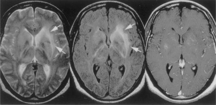
Figure 6.

Patient with African trypanosomiasis. Axial T2-weighted image (left) shows diffuse hyperintensity in both basal ganglia and along the internal, external, and extreme capsules (arrows). Axial FLAIR sequence (center, arrows) shows bilateral increased signal intensity involving the internal and external capsules bilaterally. Axial T1-weighted gadolinium-enhanced image (right) shows minimal enhancement in the basal ganglia bilaterally. (Reprinted with permission from Gill DS, Chatha DS, del Cardio-O’Donovan R. MR imaging findings in African trypansomiasis. AJNR Am J Neuroradiol 2003;24:1383–1385. Copyright # by American Society of Neuroradiology, www.ajnr.org.)
