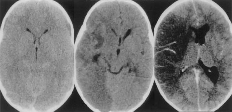
Figure 7.

Patient with Naegleria fowlerii infection identified postmortem. Noncontrast CT (left) at the level of the superior colliculus showing effacement of the quadrigeminal plate cistern. Noncontrast CT in middle shows low attenuation in the right hemisphere with mass effect. Contrast infusion 1 week later (right) shows mild vascular enhancement and persistent low attenuation consistent with infarction of both middle cerebral and posterior cerebral artery territories. (Reprinted with permission from Schumacher DJ, Tien RD, Lane K. Neuroimaging findings in rare amebic infections of the central nervous system. AJNR Am J Neuroradiol 1995;16(suppl):930–935. Copyright # by American Society of Neuroradiology, www.ajnr.org.)
