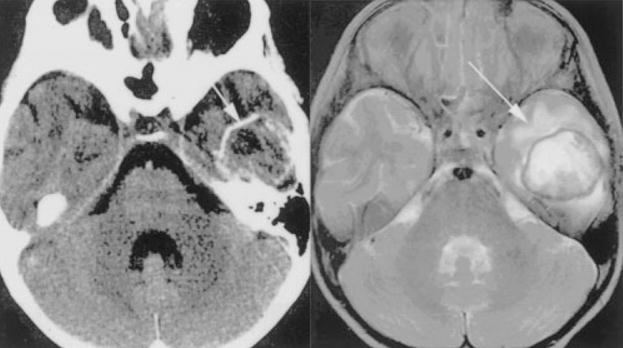
Figure 8.

Balamuthia mandrillaris infection in a 5-year-old immunocompetent girl. Head CT with contrast (left) demonstrates a ring-enhancing lesion in the left temporal lobe and T2 MRI (right) confirms the presence of edema surrounding the well-demarcated solitary abscess (arrows). (Reprinted with permission from Healy JF. Balamuthia amebic encephalitis: radiographic and pathologic findings. AJNR Am J Neuroradiol 2002;23:486–489. Copyright # by American Society of Neuroradiology, www.ajnr.org.)
