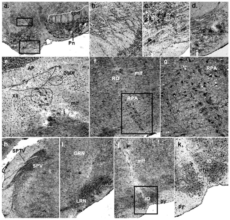
Fig. 9.
Immunolocalization of mPMAT protein in the midbrain and hindbrain. (a–d) Dense mPMAT staining in the pontine nuclei cell bodies (a, c, d) and fibers (a, b). (e) Expression of mPMAT in the medulla showing labeling of cells in the area postrema, dorsal motor nucleus of the vagus nerve, hypoglossal nucleus and medial longitudinal fasciculus. (f, g) mPMAT immunoreactivity in neurons of the nucleus raphe obscurus and nucleus raphe pallidus. Panel g contains the boxed region in panel f. (h–k) Immunolabeling of the spinal trigeminal nerve tract and nucleus, gigantoreticular and lateral reticular nucleus and inferior olivary complex. Images were obtained at 20× (a, e, h–j) and 40× (b–d, f, g, k) magnification.

