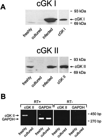
Figure 2.
Analysis of endogenous and reexpressed cGK I and cGK II in CNT/CCD cells. (A) Immunoblots of samples (20 μg protein each) from homogenates of either freshly immunodissected CNT/CCD tubules, monolayers of cells cultured from these tubules, or monolayers infected with adenoviral-cGK constructs (described in Materials and Methods) were labeled with antibodies against cGK I or cGK II (A). Shown in the right lane of each blot are standards (2 ng) of either purified bovine lung cGK I or recombinant rat intestine cGK II. The 86-kDa protein endogenously present in freshly isolated cells was identified as cGK II, and this was confirmed with immunoblots by using an additional antibody independently raised against cGK II (data not shown). (B) cGK II mRNA was detected by RT-PCR in freshly isolated and cultured CNT/CCD cells. Samples of RT-PCR-derived cGK II (420 bp) and glyceraldehyde-3-phosphate dehydrogenase (309 bp) cDNA were analyzed on an ethidium bromide-stained 2% (wt/vol) agarose gel. PCR was carried out either with (RT+) or without (RT−) reverse transcriptase. RNA (2 μg) was used as starting material for RT-PCRs.

