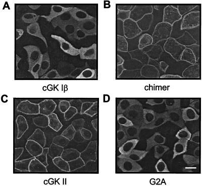
Figure 4.
Subcellular localization of cGK proteins expressed in CNT/CCD monolayers. Monolayers were infected with adenovirus for expressing either cGK Iβ (A), a chimer of the N terminus of cGK II linked to the N terminus of full-length cGK Iβ (B), cGK II (C), or a myristoylation-deficient (G2A) cGK II mutant (D). Two days after infection, immunolocalization of the cGK proteins was visualized by confocal laser-scanning microscopy. Membrane-associated (B and C) or cytosolic (A and D) localization was observed. Specificity of cGK immunostaining was confirmed by the absence of signal in noninfected monolayers (not shown). (Bar = 5 μm.)

