
Figure 3. CPI expression in Ad-VEGF-A164 injected ears.
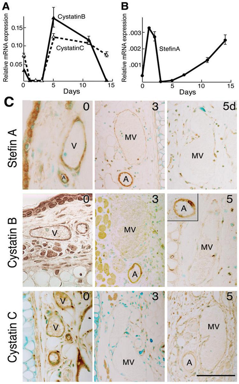
(A, B) Quantitative RT-PCR demonstrating CPI mRNA expression patterns at indicated times after Ad-VEGFA164 injection (mean ± SD). Representative data from 9 different experiments performed on 7 different animals.
(C) Immunohistochemical staining of CPIs in control ears (time 0) and at 3 and 5 days after Ad-VEGF-A164 injection. Staining for all three CPIs is greatly reduced in mother vessels (MV) compared with normal venules (V). Arteriole (A) staining for cystatin C, but not for cystatin B or stefin A, was also reduced following Ad-VEGF-A164 injection. Scale bar, 50μm.