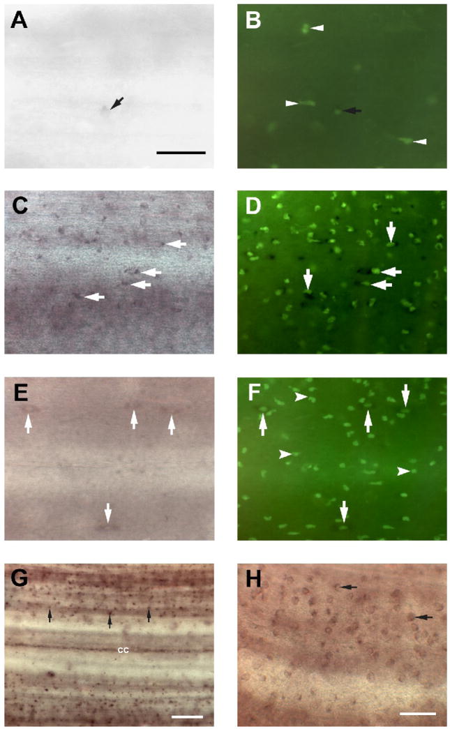
Figure 3. Microglial activation after spinal cord transection includes RGM upregulation.
A, In situ hybridization in control spinal cord wholemount revealed the presence of small RGM-expressing cells (black arrow) on the surface of the spinal cord. Scale bar, 20 μm. B, Fluorescein-labeled GSL I–isolectin B4 histochemistry revealed several lectin-labeled cells in control spinal cord (white arrowheads) including RGM–expressing cells (black arrow). In controls, few isolectin B4-labeled profiles were RGM-positive. C, Two weeks after spinal cord transection, increased numbers of small, rounded cells (white arrows), reminiscent of activated microglia/macrophages, were labeled with the RGM probe, and a dense accumulation of reactive macrophages/microglial cells, as evidenced by increasing IB4 lectin reactivity, was seen (D). Most of the lectin-labeled cells co-expressed RGM mRNA (white arrows). E, Only a few RGM–expressing cells were detected in the spinal cord 1 month after injury. Some slender, elongated RGM-expressing cells (white arrows) resembled the macrophages/microglial cells seen in control spinal cord (Fig. 3A). F, At this time, numerous small, round macrophages/microglial cells (white arrowheads) could be detected by labeling with IB4 lectin in the spinal cord, but these were never labeled by the RGM probe. On the other hand, there were some larger, elongated RGM-expressing cells that also were labeled with IB4 lectin (white arrows). G and H, RGM-expressing cells exhibited very small rounded cell bodies (no larger than 5 μm) and possessed obvious non-neuronal morphology (black arrows). Scale bar, 20 μm(G, H).

