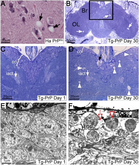
Figure 2. PrP induces spongiform degeneration of brain neurons.
(A) The brain of a scrapie-infected hamster (Ha PrPSc) contains vacuolated neurons in the medulla (arrows). (B–D) The brain of a 1 day-old Tg-PrP fly (da-Gal4/PrP-M6) displays a well-preserved neuropile and a thick layer of cells in the cortex (co) (C). In contrast, the brain of a 30 day-old flies contain abundant vacuoles in the brain (Br) and the optic lobes (OL) (B, arrows). The boxed area (magnified in D) shows prominent vacuolar degeneration of the neuropile (arrowheads) and the cortex (black arrow), and the cortex is thinner. The inner antennocerebral tract (iact, white arrow in C and D) is shown as a landmark for section depth. (E and F) Electron micrographs of brain neurons from 1 (E) or 40 day-old (F) Tg-PrP flies. 1 day-old flies exhibit normal cellular morphology (nu = nucleus), but older flies show vacuolated cytoplasm (arrow), nuclear condensation (arrowhead) and abnormal membrane folds.

